Today, dental implants are one of the most preferred treatment methods to replace lost teeth. However, a detailed diagnosis is necessary to make proper treatment planning. At this point, CBCT screening for dental implants is quite important.
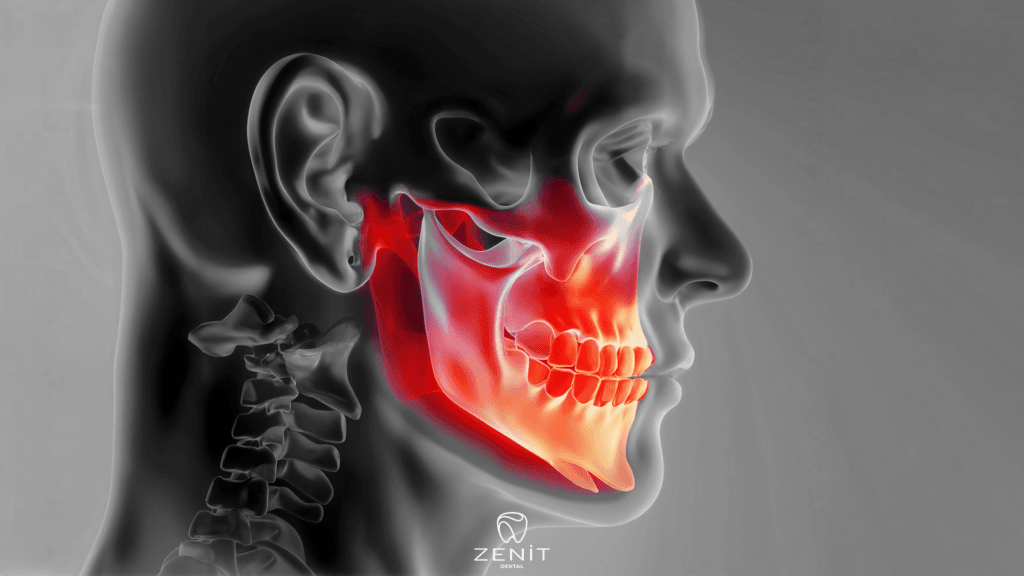
CBCT is an imaging technique for dental implants that provides more detailed images than traditional X-rays. The CBCT process produces detailed images of teeth, bone structure and sinuses. These images provide all the information needed for the correct placement of the implant.
CBCT scanning can be used to determine the size, shape and position of the implant. In addition, the scan results also show the quality and density of bone in the area where the implant will be placed. This information helps determine how stable the implant will be and whether it will integrate successfully.
CBCT screening for dental implants can also be used to facilitate surgical planning. The scan results are used to determine where to place the implant, at what angle to place it, and how deep to place the implant. This information makes the operation more precise and successful.
What is Dental CBCT?
Dental CBCT is an imaging method often used in the field of dentistry. CBCT stands for “Cone Beam Computed Tomography”. The dental CBCT process is used to obtain three-dimensional images. Dentists turn to dental CBCT scans to examine the tooth and jaw structures of their patients.
The CBCT scan creates a 3D image of teeth, bone structure and sinuses. This method helps dentists in assessing the placement of teeth and the health of surrounding tissues. It can also be used for implant procedures. Dental Cone Beam Computed Tomography scanning, like other imaging methods, involves radiation, but the amount of radiation exposed is quite low.
Dental scanning provides more detailed images compared to other imaging methods used in the field of dentistry. For this reason, it is often used by dentists to make an accurate diagnosis and treatment plan. However, the cost of this method is higher than other methods.
CBCT screening has become a rapidly becoming a widespread imaging method in the field of dentistry. This method allows dentists to obtain a three-dimensional image of the teeth and jaw structures. This provides a more accurate and effective way to diagnose and treat patients.
How is Dental CBCT Done?

Dental CBCT (Cone Beam Computed Tomography) is a screening method widely used in the field of dentistry today. This method allows to view the internal structure of the teeth and gums in detail. Dentists may perform CBCT screening to diagnose their patients’ oral health or to schedule surgical intervention. So, how is dental CBCT done?
Dental Cone Beam Computed Tomography scanning is a technology used to extract a detailed 3D image of the upper and lower parts of the mouth. This screening is prescribed by the dentist and done by the radiology technician. The screening process is carried out in a device similar to a dentist’s chair. This device scans the patient’s head and jaw by keeping them steady.
Unlike other radiological scanning methods, this process creates a three-dimensional image instead of individual sections. During the scan, X-rays rotate with focus on the patient’s mouth. The data received during this rotation is processed on a computer and a three-dimensional digital model is created. By looking at this model, the dentist can analyze in detail the internal structure of the patient’s teeth and gums.
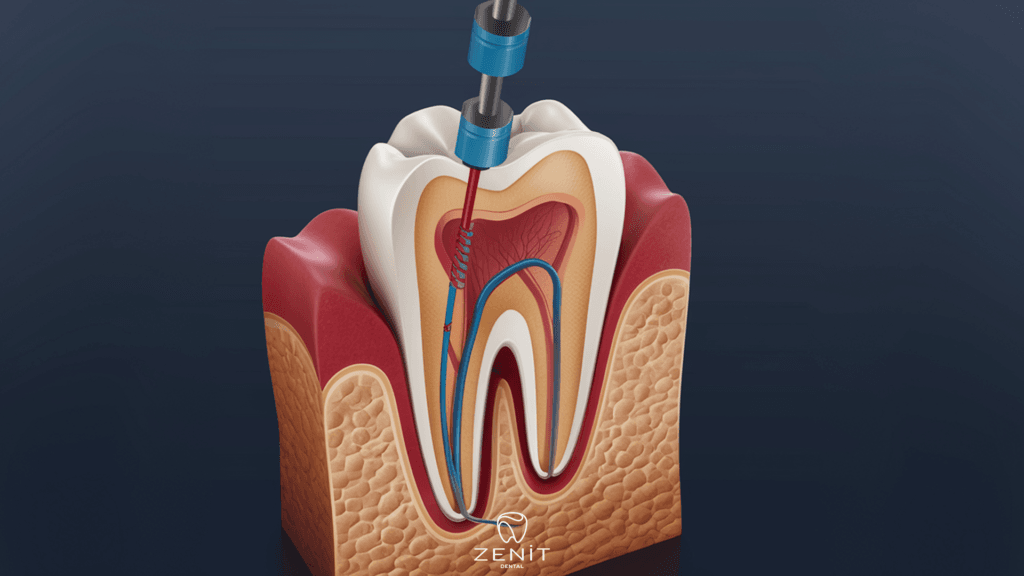
Dental CBCT is used to detect intraoral cysts, tumors, tooth inflammation, sinus problems and problems in bone structures. With this screening, it becomes possible for patients to intervene early with previously undetectable dental problems.
Is CBCT Used for Implant Surgery?
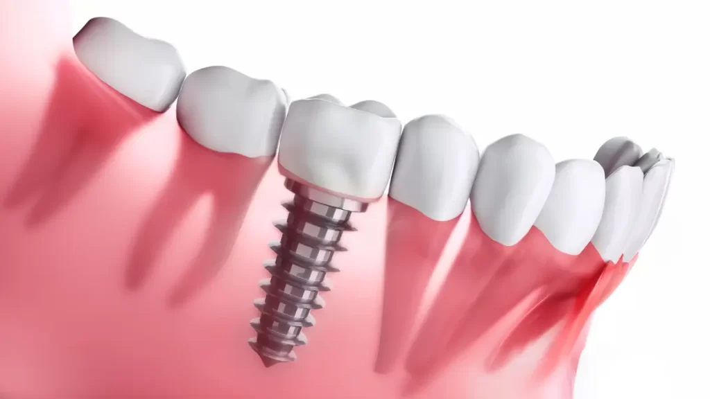
Implant surgery is one of the most effective methods used to treat missing teeth. This process places artificial tooth roots on your teeth, making your teeth look more robust and natural. But for implant surgery, an accurate diagnosis is necessary to fully understand the condition of your teeth. For this reason, dentists often use CBCT scans.
CBCT is an imaging method used by dentists to examine the condition of a patient’s teeth before implant surgery. This method is the digital X-ray technique used by dentists to obtain three-dimensional images.
CBCT scans allow dentists to view your teeth from a 360-degree angle. In this way, a more accurate diagnosis can be made about the condition of your teeth before implant surgery. CBCT scans are used to determine the bone structure of your teeth, the location of nerves, the size of the area where the implant will be placed, and the overall condition of your teeth.
CBCT screening is very important for dentists before implant surgery. Because of these scans, dentists can determine if you are suitable for implant surgery. In addition, CBCT scans also allow dentists to perform procedures more accurately and precisely during implant surgery.
What is the Role of CBCT in Implant Dentistry?
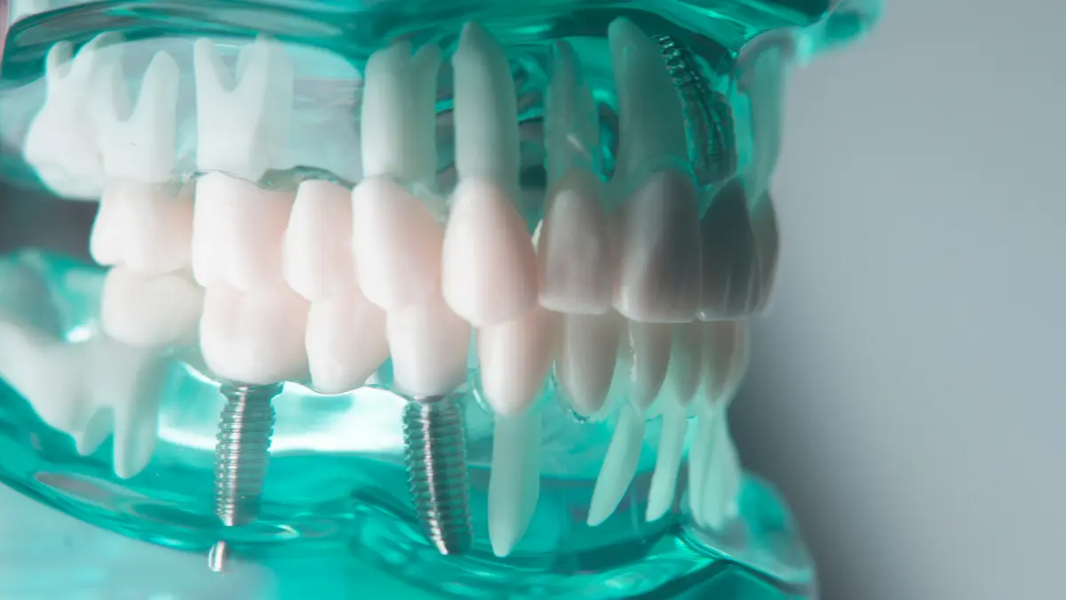
Tooth loss is a very common problem today. Many dentists find a solution to this problem with implant dentistry. However, regardless of the cause of tooth deficiency, it is extremely important to make the correct diagnosis and planning for implant tooth treatment. At this point, Cone Beam Computed Tomography imaging technology plays a big role in implant dentistry.

CBCT is a type of CT scanning technology that provides 3D imaging. The use of this technology is very important to make the correct diagnosis and planning in implant dental treatment. Traditional X-ray technologies provide only two-dimensional imaging. In addition, while it shows an image of only one area, Cone Beam Computed Tomographytechnology shows all the surrounding tooth and jaw structures in detail.
These images allow the dentist to make a much more detailed diagnosis prior to implant dentistry. It helps to complete the tooth structure before the implant is placed. Thus, it ensures that the implant is placed in the right place and at the right depth. It also reduces the risk of damaging neighboring teeth and sinuses when placing the implant.
Cone Beam Computed Tomography also increases the success rate of implant dental treatment. Before the implant is placed, the dentist can make a detailed evaluation of the patient’s jaw structure and bone density. This ensures that the implants are selected in the right size and shape and that they are placed appropriately. This causes the implants to last longer and have fewer complications.